

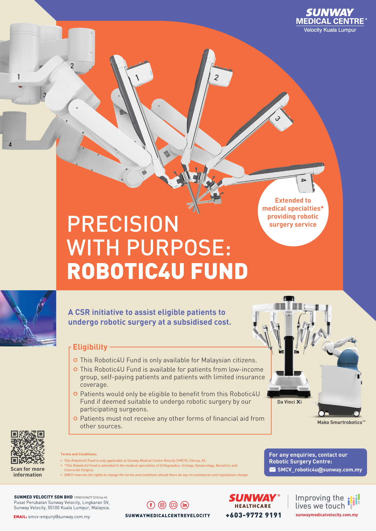
Our Radiology department provides clinical services in diagnostic radiology, interventional radiology, ultrasound and cross sectional imaging (CT scan and magnetic resonance imaging) with our state-of-the-art equipment and facilities.
Services and Procedures offered:
1. 3T MRI Scanner
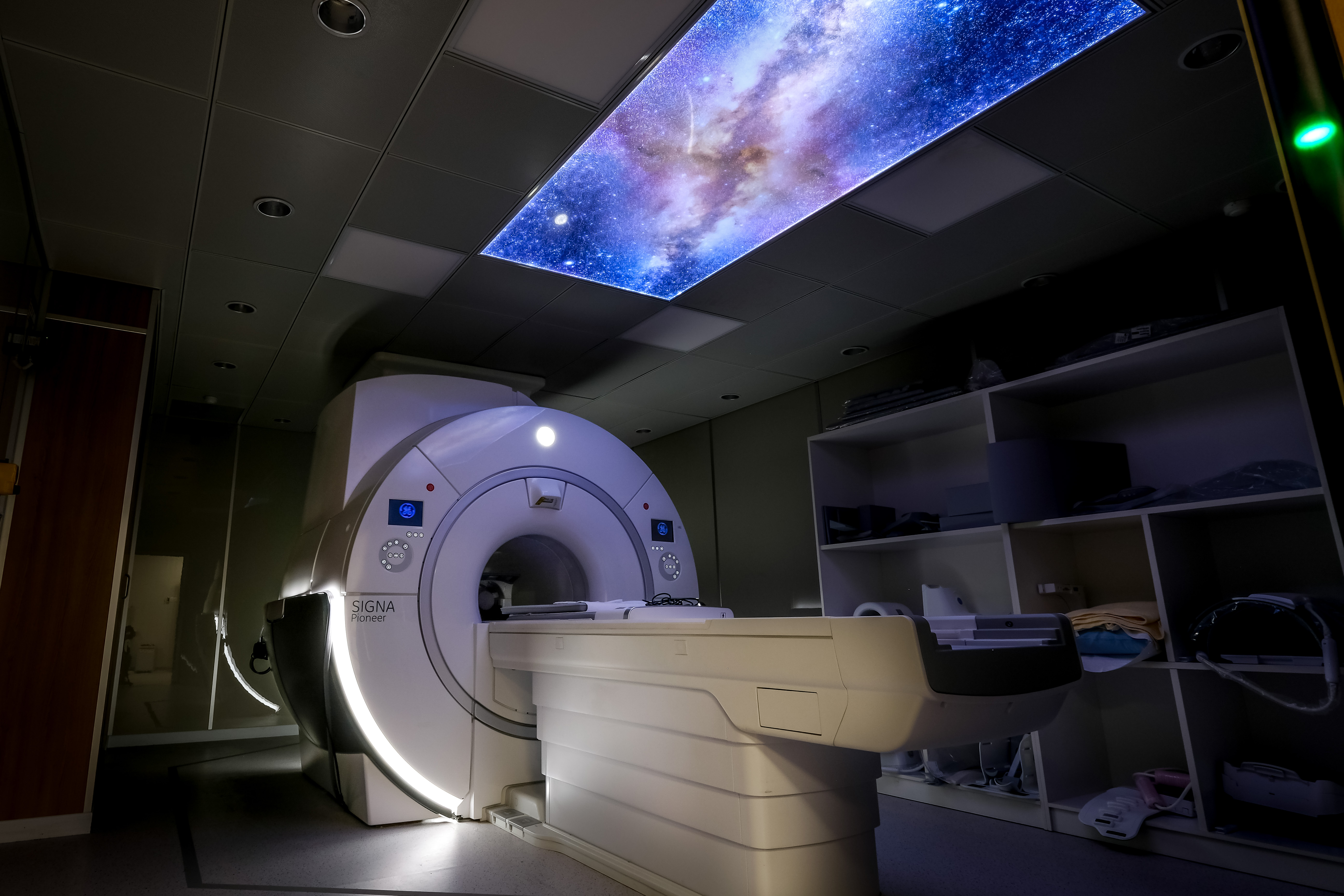
The 3 Tesla (3T) MRI is an advanced magnetic resonance imaging (MRI) scanner with twice the field strength of most conventional MRI scanners. It allows for faster scans, greater detail, clearer images, and more accurate diagnoses.
3 Tesla (3T) MRI is particularly useful in scans of the brain and spinal column, and of the musculoskeletal system.
Some of the procedures that the 3 Tesla (3T) MRI will benefit include:
i. MR angiography
ii. Neurological/brain imaging
iii. Spine studies
iv. Orthopedic – including elbow, wrist, hip, knee, foot and ankle
v. Prostate
vi. Pelvis – Male and Female
vii. Abdominal
viii. Functional imaging, spectroscopy, and brain fiber tracking
ix. Breast MRI
What to expect during your 3 Tesla (3T) MRI test:
During an MRI scan, you will need to remain very still to avoid blurriness in the scanned images. Depending on your condition, monitors may be used to track your pulse, heart rate, and breathing.
If a contrast dye is used, a small IV needle is inserted into your hand or arm and a saline solution is dripped into your vein to prevent clotting before the dye is injected.
Next, the technologist will leave the room but will continue to communicate with you through an intercom. The technologist will give you directions, such as to hold your breath or remain still. You will then hear clicking sounds as pictures are taken by the technologist. When the test is done, you will slide out of the scanner.
Your scans will then be interpreted by a radiologist and delivered to your consulting doctor.
2. 256 slice CT Scanner
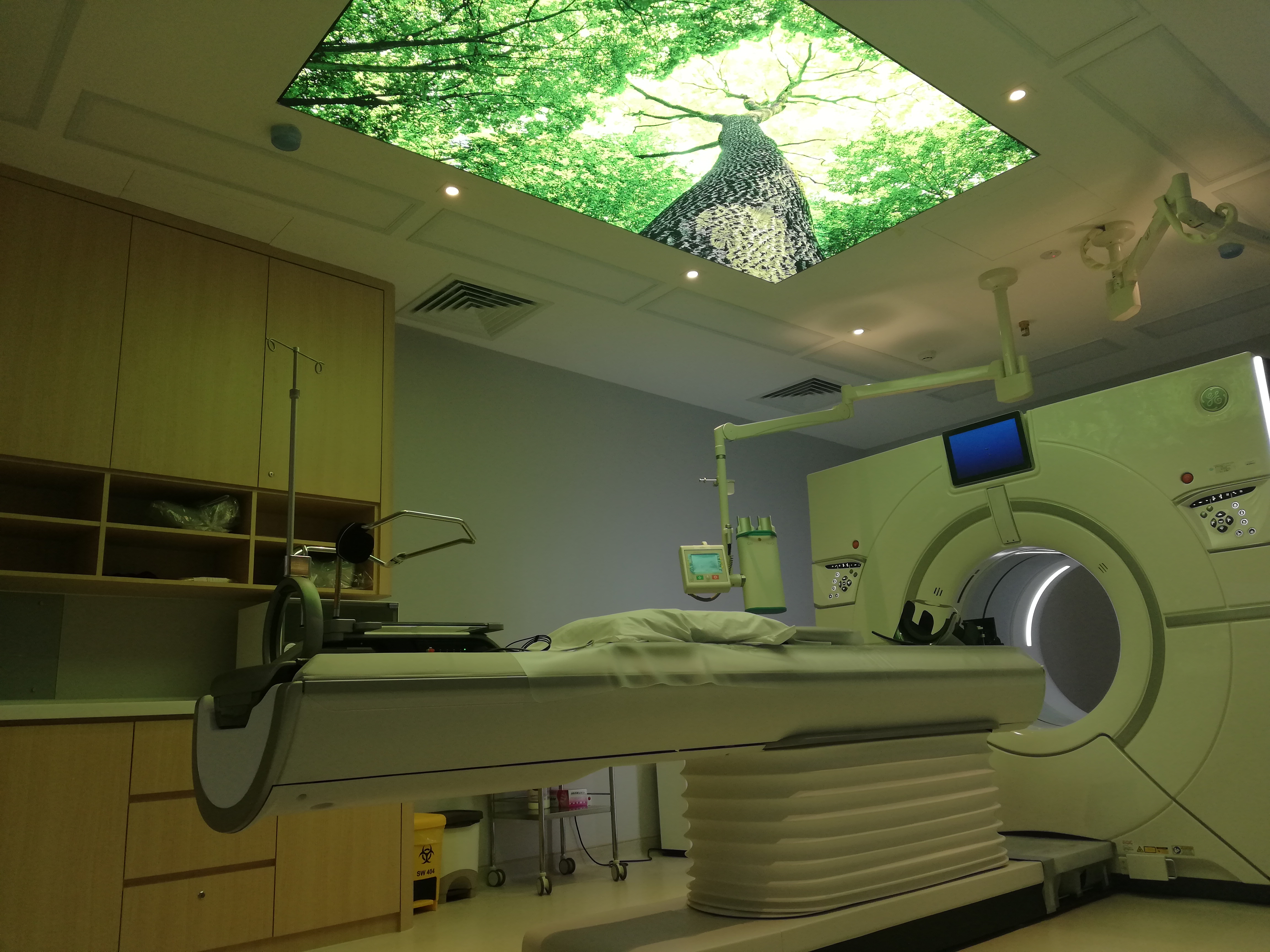
With the latest technology and advance post processing software, our 256 slice CT Scanner capable to produce high quality images with lower radiation dose compare to conventional CT Scanner. A versatile scanner for:
- Emergency Care
- Oncology
- Stroke
- MSK
- Pediatrics
- Cardiac
- Low dose CT Lung
What to expect during your CT Scan
Before a CT scan, let your doctor know if:
- You have ever had a reaction to any contrast dye, or if you are allergic to iodine or seafood
- You are pregnant or think you may be pregnant
- You are claustrophobic or tend to become anxious easily
You will be asked to remove any clothing, jewellery or objects that could interfere with the scan. If a contrast is used, it will be administered through an IV or given as oral medication to swallow. You will need to remain still and quiet during procedure which may last 10 to 20 minutes. During the test, you will be able to communicate with the technologist through an intercom.
An experienced radiologist will then interpret your scans and deliver them to your consulting doctor.
3. 3D Mammogram
Mammography is a specific type of imaging that uses a low-dose x-ray system to examine breasts. Both digital and conventional mammography use x-rays to produce an image of the breast. However, in digital mammography, the image is stored as a computer file (instead of a piece of film) making it easier for the image to be enhanced, magnified and retrieved for further evaluation.
A mammogram is used to detect and diagnose breast disease in women experiencing symptoms such as a lump, pain or nipple discharge. Even when a woman experiences no symptoms, a mammogram is an important screening tool used to detect early breast cancer. Women above the age of 40 are encouraged to get a mammogram every year as this test can discover a lump up to two years before it can be felt.
How to prepare for a digital mammography
The procedure for having a digital mammogram is the same as with conventional mammography.
When scheduling a mammogram with your doctor, be sure to discuss any new findings or problems in your breasts and inform your doctor of your family or personal history of breast cancer.
The best time for a mammogram is one week following your period. Be sure to inform your doctor or the radiographer if you think you could be pregnant. Before the mammogram, let the technologist performing the exam know of any breast symptoms you may have. If you have had previous mammograms done, it would be helpful to make them available to the radiologist at the time of the current exam
4. Bone Mineral Density
Osteoporosis is a potentially painful and crippling disease which can affect anyone but occurs more commonly in older women.
It involves the gradual loss of calcium, causing the bones to become porous, brittle and break easily. Half of all menopausal women have or are at high risk of developing osteoporosis.
Often called a silent disease, osteoporosis doesn’t produce symptoms until a fracture occurs. The cumulative life-time fracture risk for a 50-year-old woman with osteoporosis is as high as 53%.
The bones most likely to break are the hip, spine and forearm.
Bone density scans are used to diagnose osteoporosis and are recommended if you:
- are a thin and tall menopausal woman and/or not taking estrogen
- have a personal and/or maternal history of hip fracture
- milk intolerant and/or have low calcium and vitamin D intake
- a smoker or drink alcohol in excess
- are a man with clinical conditions associated with bone loss
- have a thyroid condition
- have experienced a fracture after only a mild trauma
- use medications that are known to cause bone loss, such as corticosteroids
Services & Procedures Offered
Bone density scanning is also called dual-energy x-ray absorptiometry (DXA or DEXA) or bone densitometry. It is an enhanced form of x-ray technology used to measure bone loss and is most often used to diagnose osteoporosis and assess an individual’s fracture risk. It is also used to monitor the effectiveness of your treatment. The scan is a safe, fast and painless procedure often performed on the lower spine and hip. The procedure is non-invasive, does not need anesthesia and is free from uncomfortable side-effects.
Imaging applications:
- AP lumbar spine
- hip joint
- forearm
- IVA (vertebral assessment)
Total body scan
- General and mobile Digital X-ray
Brief description will be shared soon
- Diagnostic ultrasound
Brief description will be shared soon
- CardioVascular Angiosystem (located at 6th floor Cathlab)
Brief description will be shared soon
Contact Info
For more information or to make an appointment, please call us at:
| Tel | +603 - 9772 8278 |
| Fax | N/A |
| Operation Hours |
Monday – Friday: 8.30am – 5.00pm Saturday: 8.30am – 1:00pm |
